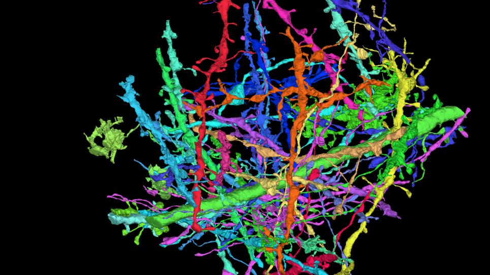
Shapes of neurons can be reconstructed from electron microscopy image data of human tissue samples. The images are analyzed using a machine learning approach to identify (segment) the neurons. In initial runs on Aurora, the team has segmented a small fraction (300 gigavoxels) of a petabyte dataset using 512 nodes. This image shows the 36 largest neuronal objects out of four million in a selected subvolume, each consisting of at least five million voxels. Note the 3um scale bar at lower left.